“Age does not bring you wisdom— age brings you wrinkles.” —Estelle Getty
As bad as one might look in a passport or driver’s license photo, nothing is scarier than the output of a modern skin imaging analysis device. To see lines and wrinkles, UV damage and red blotches, pores and excess sebum in vivid detail is enough to make anyone 20 and older run to the nearest antiaging serum in a panic. The combination of high-end digital cameras and sophisticated computer programs has made these devices available everywhere—from research labs and dermatologist’s offices to upscale cosmetic counters. A day in the sun in 1971 has left traces that can still be photographed in vivid colors, and a line you thought no one could notice now looks like the Grand Canyon.
Analysis of the skin using still or video cameras and digital image analysis (DIA) software has long been used by the medical profession to detect dangerous conditions like melanoma using parameters such as color, shape, border qualities and structural components. And Red, Green, Blue (RGB) analysis has been used for color-based recognition algorithms for many years.
Russian State Medical University researchers1 suggested using skin health indexes as indicators of aging. They evaluated skin fluorescence after being exposed to near UV radiation (365 nm [nanometers]), and estimated age-specific changes by measuring fingertip fluorescence. Experiments showed that skin fluorescence increase correlates well with chronological age of 20- to 70-year-old people.
Formation of glycotoxins (cross-linked polymers) in the dermis is considered to be one of the main reasons for skin age-specific changes and fluorescence fluctuations.
Skin Type
The first step to analyzing an individual’s skin is assigning a skin type. The most common standard is the Fitzpatrick Classification Scale, which categorizes skin from I to VII based on genetic disposition, reaction to sun exposure and tanning habits. This information is critical to correctly assessing the current condition of the skin and for recommending a treatment regimen. Estheticians are trained to do this by visual examination and a standard questionnaire. Alternatively, optical imaging devices can be programmed to assign a category automatically.
Instrumental analysis of the skin depends on the interaction of light with various molecular elements in the upper layers. Entering the skin, light undergoes wavelength-dependent absorption and scattering that provide the raw data for optical imaging. The two most important skin chromophores in the visible range of the spectrum are hemoglobin and melanin.
The skin also contains fluorophores (a component of a molecule that causes a molecule to be fluorescent) that can provide fluorescence contrast that adds an additional dimension to the image. Skin fluorpohores include collagen and elastin. Biological processes modulate the fluorescence signals in predictable ways. Such cases include intrinsic aging (See “A Closer Look at Intrinsic Aging” from the April 2009 issue of GCI magazine, also available online.), photoaging and conditions such as psoriasis, acne and non-melanoma skin cancer.
Skin analysis uses multiple light sources, normal white light, polarized light and a safe wavelength UV light (approximately 100mn), combined with a high resolution digital camera. A decade ago the Japanese microscope manufacture Scalar developed a high resolution camera that magnifies skin 160 times and was attached to an early version of skin analysis software. The result was Skin Xpert, which provided moisture levels, oil reading, elasticity and pore size measurements.
Many companies provide systems for medical and cosmetic applications, such as the Image Pro II from Enhanced Image Technologies and the Reveal Imager and Omnia Imaging System from Canfield Scientific2. The BTBP Clarity Pro from BrighTex Bio-Photonics3 is another widely used unit, and the company kindly provided images of the author to illustrate some of the information that can be teased from high definition digital images coupled with sophisticated software.
According to Francis Friscia of International Research Services, “Quantifiable scientific validation is considered the gold standard of claim substantiation. The Clarity 2 Pro R&D bioinstrumentation allows us to supply that for over 25 skin parameters.” This columnist put his face to the test, getting an analysis of wrinkles, pigmentation and UV damage.
By itself, the high resolution camera shows scary details when enlarged, but it is nothing compared to UV images, redness report or pore analysis. Fortunately, at the end of the process is a “treated skin” photo which sheds decades off the subject’s face. The treated skin photo is where “hope in a jar” finds its source.
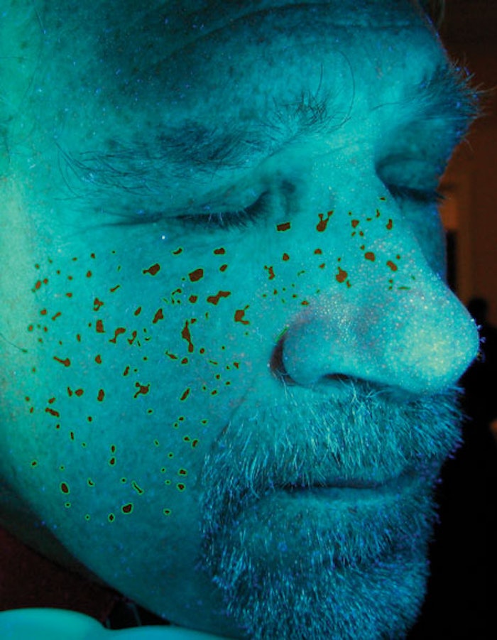
The skin parameters given qualitative form depend on brand and model: some basic categories are UV damage, complexion, skin radiance, redness (subsurface erythema), pigment, wrinkles, pores and acne. Photo 1 shows wrinkles with superimposed blue lines, Photo 2 is a pigment analysis. To achieve these images, five photos were taken with different light parameters and the results teased out with proprietary software. It is possible to have a specific program focused on lips: surface area, irregularity and definition of the vermilion border, horizontal and vertical lines. The vermilion border is the juncture between the lighter skin of the face and the redder tissue (vermilion) that is commonly called the lip. The vermilion is red because the skin is thin and there are numerous small blood vessels underneath it. A moustache, such as the authors’, will make an accurate measurement of the vermilion border impossible.
Besides producing a straightforward view of the face, eyes or lips, computer analysis can generate a 3-D model of the skin. This allows viewing skin texture and the depth and size of wrinkles in a visually compelling way.
Advanced research models contribute to medical diagnosis as well as establishing the efficacy of cosmeceuticals. The most refined medical models can analyze eight narrow band images from 400 nm to 1000 nm, combined with a complex computer algorithm to provide clinically significant data.
The image analyzer doesn’t replace other clinical tests, but it is the most visually powerful depiction of many common skin conditions. Placing a basic model on a cosmetic counter allows a consumer to see in vivid detail the effects of sun and the ravages of time, and certainly motivates the sales of treatment regimens. The ability to produce before-and-after images is a tremendous marketing tool. The analysis is noninvasive, yields quick results and can detect conditions invisible to the naked eye.
Author Acknowledgements
Thanks to Francis Friscia of IRSI and Raj Chhibber and Shefali Sharma of BrighTex Bio-Photonics for providing valuable information, images and analysis with the Clarity Pro.
REFERENCES
Steve Herman is president of Diffusion LLC, a consulting company specializing in regulatory issues, intellectual property, and technology development and transfer. He is a principal in PJS Partners, offering formulation, marketing and technology solutions for the personal care and fragrance industry. He is an adjunct professor in the Fairleigh Dickinson University Masters in Cosmetic Science program and is a Fellow in the Society of Cosmetic Chemists.